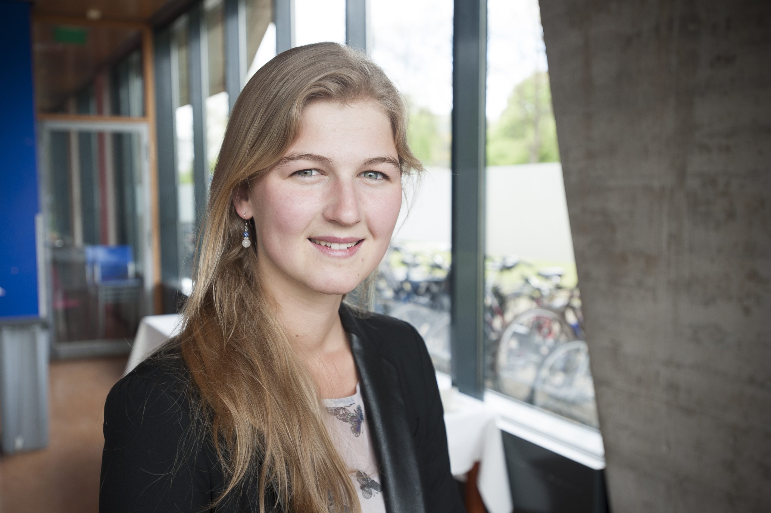
Master’s student Inès Beekers (applied physics) was awarded the second prize of the Dutch Physics Society (NNV) for her bachelor’s thesis on scanning tumours with ultrasound.
Three students presented their BSc. thesis at the FYSICA 2014 final in Leiden on April 1 2014. They were Melissa van Beekveld (University Utrecht; supersymmetry), Inès Beekers (TU Delft; ultrasound) and Jonas Voorzanger (VU A’dam; photoconductivity).
Melissa van Beekveld won the jury’s hearts with her enthusiastic presentation on the abstract concept ‘supersymmetry’. By making plausible that supersymmetry (the proposition that each boson has a fermion as its superpartner) may be hidden in the existing data, she won the first prize.
The second prize was awarded to TU’s Inès Beekers about whom the jury wrote: ‘She chose a subject with societal impact: early diagnosis of cancer with ultrasound. The jury praises her creative and many-sided input in the project.’
Her BSc supervisor Dr. Martin Verweij at Applied Sciences is equally laudatory: “She has analysed the task and proceeded from there. Both her theoretical and hands-on skills were very good. She made a phantom for measurements, which she carried through. Her work could well have passed for a master’s thesis.” Verweij rated her thesis with a 9.5
Beekers says she wanted to do a useful physics project, which led her to doing a minor in medicine at the Erasmus Medical Centre. Subsequently she contacted Verweij from Acoustical Wavefield Imaging (faculty Applied Sciences) who suggested researching the use of ultrasound for imaging tumours. Considering that ultrasound is a ubiquitous tool in hospitals, being able to use them for tumour testing would seem a welcome idea.
Tumours have characteristic chaotic patterns of blood vessels. In normal tissue blood vessels mainly run parallel. But in a tumour they crisscross the tissue almost at random. So if one could image the direction of these vessels, you might be able to identify a tumour from surrounding tissue.
Trouble is: these blood vessels are typically 0.01 – 0.1 millimetres in diameter and ultrasound resolution is in the order of a millimetre. So, can this resolution gap be bridged?

Imaging blood is greatly enhanced by adding encapsulated micron-sized bubbles to the flow: up to ten million per millilitre. In clinical practice these bubbles with a lipid shell are known as ‘contrast agent’. They make blood vessels light up under the ultrasound scan.
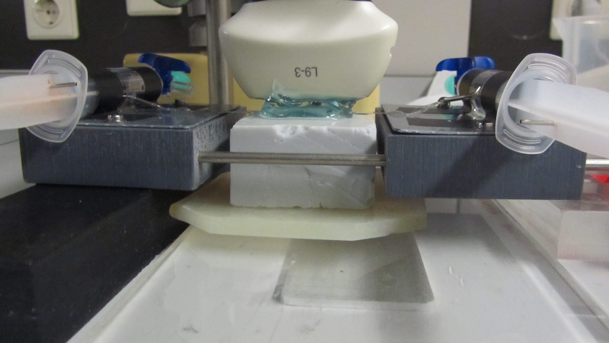
In the lab, Beekers made a phantom with ten very fine tubes 0,2 millimetre across and as near to each other as possible. She gently forced a saline solution through them carrying lipide bubbles for contrast. And much to her own surprise, she could detect flow through the tubes (which modeled blood vessels) although the distance between them is five to ten times smaller than the nominal resolution of the ultrasound scanner.
With her phantom featuring ten stretched tubes, Beekers showed that fine flows could be observed if they align with the direction of the ultrasound sweep scan, but hardly so if the flow is perpendicular to the scan. This may be key for distinguishing tumours from healthy tissue.
“This is all still very fundamental”, Beekers stresses. Nonetheless, the idea is that you could identify a tumour on a suspect spot by performing two perpendicular scans. Healthy tissue would give ‘polarised’ readings: maximum flow in one direction and almost nothing under a right angle. A tumour, due to its crisscrossed vessels, would show much less difference in the two perpendicular readings.
The finding is as yet too fundamental for the industry to pick it up, Beekers thinks. The next step would involve testing with a more realistic 3D phantom to see if the polarisation effect manifests itself.
Such a project isn’t planned yet, says Verweij. But it’s in his head together with numerous other plans he discusses with students interested to do a project in medical imaging.
→ Dalien Inès Beekers, Identification of tumor vasculature with ultrasound imaging, BSc. thesis supervisor Dr. Martin Verweij (AS)


Comments are closed.