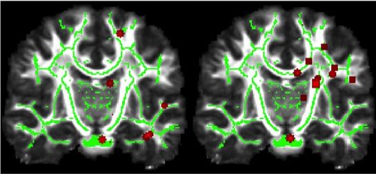
Changes in the structure of the brain in people developing Alzheimer’s disease and Amyotrophic lateral sclerosis (ALS) could be monitored more precisely thanks to new research at TU Delft.
Magnetic Resonance Imaging (MRI) is a technique that uses magnetic fields and radio waves to take pictures of the organs of the body; Diffusion Imaging is a type of MRI that reveals the microscopic structure of tissues within the organs. Dr Frans Vos, Associate Professor in Quantitative Imaging Group, Department of Imaging Physics at TU Delft said, “There are two types of matter in the brain: grey matter which is the thinking bit, and white matter which is made of connective fibres. Diffusion Imaging is often used by neurosurgeons to help them avoid cutting through white matter when removing a tumour for instance.”
Diffusion Imaging is also used to detect changes in the structure of the white matter, which is what happens in neurodegenerative diseases such as ALS and Alzheimer’s disease. “Ideally we’d like to be able to quantify these changes”, added Vos, “so measure the change in structural integrity of the white matter.” One of Vos’ students, Jianfei Yang, has been doing just this, obtaining his PhD (Analysis of Diffusion MRI: Disentangling the Entangled Brain) in the process.
Ink spots
Diffusion Imaging compares patterns produced by water molecules moving within the body’s cells. “If you drip ink on to tissue paper,” explained Yang, “the spot will be circular. But if you drip ink on to newspaper, the spot will be elliptical. That’s because newspaper is made up of fibres stretched in one particular direction and the ink will move along those fibres, producing an ellipse.” The same principle applies to Diffusion Imaging of the white matter: in healthy white matter, water molecules diffuse along the fibres, which are covered in a protective sheath, producing an elliptical pattern. But if a person has a neurodegenerative disease, the fibres are breaking down and water molecules not only move along the fibres but also in other directions, producing a rounder diffusion pattern.
“What Jianfei has done,” explained Vos, “is help distinguish whether there are one, two or three tracts of white matter running in different directions, and then modelled the diffusion of each tract separately, increasing the sensitivity of the analysis.” This gives researchers a better insight into how white matter degenerates because of neurodegenerative disease.


Comments are closed.