Inspection of the intestines with a camera is the standard way to evaluate inflammatory bowel disease. A research consortium as developed a non-invasive alternative with magnetic resonance imaging (MRI)

With some 700 thousand Crohn’s disease patients in Europe alone, the societal need for this development is evident. Crohn’s disease is an inflammatory bowel disease with varying intensity, not only between patients, but in time as well. Varying levels of severity need different treatments.
Despite a large number of different methods and approaches, there is no objective, reproducible, quantitative and non-invasive method to measure the severity of the disease. In the clinic, visual inspection by colonoscopy is the standard although cross-sectional techniques such as CT and MRI are increasingly being used as well.
The European research programme VIGOR++ (Virtual Gastrointestinal Tract) aimed for a better management of the disease (which is incurable) through the development of a personalised virtual gastrointestinal model. Frans Vos (Imaging Physics at TNW Faculty) coordinated the programme in which also the abdominal radiology departments of UCL (London) and AMC (Amsterdam) participated.
The PhD research by Dr. Robiel Naziroglu was part of the VIGOR++ programme. He analysed magnetic resonance images in order to quantify the activity of Crohn’s disease. Naziroglu presented his thesis ‘Analysis methods for abdominal imaging’ on Monday November 21, 2016.
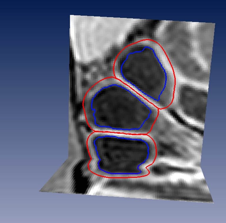
Imaging intestines by MRI is no easy feat. The varying content of the guts, narrowings and diversity of the surrounding tissues are just a few factors making it difficult to unambiguously determine the inner surface, the outer surface and features such as the bowel wall thickness.
 reproducibility
reproducibility
reproducibility
In a semi-automatic measurement technique that Naziroglu developed, the medical specialist coarsely draws a centre-line in the middle of the bowel, after which the computer identifies the outlines of the bowel. The method resulted in measurements that had a much higher correlation between various observers than the interpretations by the eye. In other words: the semi-automatic method significantly increases the reproducibility of the MRI analysis.
Next to the semi-automatic identification of the bowel, Naziroglu also developed a scoring system for the disease based on various manual and automated MRI-features. Compared to other scoring methods currently in use, the new method showed a much higher agreement between observers. So here again, the reproducibility has been improved.
Although the research was done in close collaboration with medical staff from the AMC, further developments are needed to make the new MRI analysis method practically usable by medical specialists.
Naziroglu said: “The method needs further testing and development before it can be applied in practice. Nonetheless, I think the new method is very promising.”
CV: Robiel Naiziroglu (29) was born in Utrecht. He obtained his Gymnasium diploma from the Christelijk Lyceum Zeist in 2005 and went on to study applied physics at the TU Delft. He finished his master’s in Biomedical Engineering in 2011, after which he started as a PhD candidate involved in the VIGOR++ project at the Quantitative Imaging group (Faculty of Applied Sciences)
Robiel Estifanas Naziroglu, Analysis methods for abdominal imaging: quantifying Crohn’s disease activity, PhD supervisors Prof. Lucas van Vliet (TNW), Prof. J. Stoker (AMC) and Dr. Frans Vos (TU / AMC), November 21, 2016.
Heb je een vraag of opmerking over dit artikel?
delta@tudelft.nl
Comments are closed.