Dr. Eduardo Margallo Balbás’ thesis contains enough material for at least three PhD dissertations. One of his most promising inventions is an implant that uses light to fight brain tumors.
“I wasn’t told to do anything, so I worked on many different things, looking for opportunities”, says dr. Eduardo Margallo Balbás, whose PhD program was aimed ‘simply’ at finding new research lines related to optical techniques for medical applications. It resulted in his thesis, ‘Optical techniques for the study of living tissue’, which he defended in May.
When Balbás started off four years ago, one idea led to another: a dentist drill with a built-in camera; an instrument that analyses bone structures without x-rays; and a spinal cage used to graft vertebra and cure some cases of degenerative spinal disorders. This is merely a selection of some of the instruments he developed.
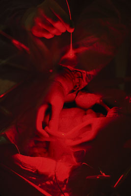
The last thing Balbás worked on, which also happens to be one of the most promising technologies, is an implant for the brain that emits light pulses. He developed this device together with researchers at the Erasmus Medical Centre. The implant should help to better eradicate brain tumors by the use of so-called ‘photodynamic therapy’.
With photodynamic therapy, photosensitive drugs inside the body, which are normally inactive, are activated by light. Using this method to treat internal organs is quite problematic, however. Such treatments are sometimes done with endoscopes and fibre optic catheters serving to deliver light. An implant would allow the patients to easily administer the light themselves at the appropriate times, simply by using a remote control.
Balbás’ instrument has generated lots of interest: the Medical Delta, a joint venture involving the universities of Leiden, Rotterdam, and Delft, recently applied for a grant to further develop this implant.
The same cannot be said for some of his earlier inventions, sophisticated as they may be. Take for instance the dentist drill with a built-in camera in the tip of the drill. Balbás smiles: “With this device, dentists can see through the bone a millimeter ahead while drilling. This should help them to avoid drilling through arteries or other delicate structures. But we’re told that for this feature dentists would only be willing to pay half the price of what the drill would cost.”
The drill uses optical coherence tomography (OCT), an interferometric technique used to create three-dimensional images from biological tissues. The technique is much used by ophthamologists to create images of the retina.
OCT-machines are normally very big because they contain mirrors, which are necessary to delay light beams. As this is hardly the thing one could put in a millimetre-sized drill tip, the Delft researcher has therefore developed a silicon chip that heats up to a 100° and then cools down again at a rate of 10,000 times per second. Balbás: “When it’s hot the light moves slower. I managed to delay the light by three picoseconds (0.000 000 000 003 seconds). Not many people thought it was possible to delay light that much.”
During his bone research, Balbás also fine-tuned a technique that might one day enable doctors to analyze bone structures with near infrared light instead of x-ray.
“Bone often consists of honeycomb hollow structures filled with marrow”, Balbás explains. “By shooting light pulses through the bone and measuring the scattering and absorption properties, you can get an idea of the amount of bone mineral and thus the strength of the bone.”
This is useful for predicting osteoporosis. Currently x-rays are used for this purpose, but these rays are harmful, so they can’t be used too often. If you want to monitor people, light might be a better option, Balbás believes.
“I wasn’t told to do anything, so I worked on many different things, looking for opportunities”, says dr. Eduardo Margallo Balbás, whose PhD program was aimed ‘simply’ at finding new research lines related to optical techniques for medical applications. It resulted in his thesis, ‘Optical techniques for the study of living tissue’, which he defended in May.
When Margallo started off four years ago, one idea led to another: a dentist drill with a built-in camera; an instrument that analyses bone structures without x-rays; and a spinal cage used to graft vertebra and cure some cases of degenerative spinal disorders. This is merely a selection of some of the instruments he developed.
The last thing Margallo worked on, which also happens to be one of the most promising technologies, is an implant for the brain that emits light pulses. He developed this device together with researchers at the Erasmus Medical Centre. The implant should help to better eradicate brain tumors by the use of so-called ‘photodynamic therapy’.
With photodynamic therapy, photosensitive drugs inside the body, which are normally inactive, are activated by light. Using this method to treat internal organs is quite problematic, however. Such treatments are sometimes done with endoscopes and fibre optic catheters serving to deliver light. An implant would allow the patients to easily administer the light themselves at the appropriate times, simply by using a remote control.
Margallo’s instrument has generated lots of interest: the Medical Delta, a joint venture involving the universities of Leiden, Rotterdam, and Delft, recently applied for a grant to further develop this implant.
The same cannot be said for some of his earlier inventions, sophisticated as they may be. Take for instance the dentist drill with a built-in camera in the tip of the drill. Margallo smiles: “With this device, dentists can see through the bone a millimeter ahead while drilling. This should help them to avoid drilling through arteries or other delicate structures. But we’re told that for this feature dentists would not be willing to pay a high price.”
The drill uses optical coherence tomography (OCT), an interferometric technique used to create three-dimensional images from biological tissues. The technique is much used by ophthamologists to create images of the retina.
OCT-machines are normally very big because they contain mirrors, which are necessary to delay light beams. As this is hardly the thing one could put in a millimetre-sized drill tip, the Delft researcher has therefore developed a silicon chip that heats up to a 100° and then cools down again at a rate of 10,000 times per second. Balbás: “When it’s hot the light moves slower. I managed to delay the light by three picoseconds (0.000 000 000 003 seconds). Not many people thought it was possible to delay light that much in this way.”
During his bone research, the scientist also fine-tuned a technique that might one day enable doctors to analyze bone structures with near infrared light instead of x-ray.
“Bone often consists of honeycomb hollow structures filled with marrow”, Margallo explains. “By shooting light pulses through the bone and measuring the scattering and absorption properties, you can get an idea of the amount of bone mineral and thus the strength of the bone.”
This is useful for predicting osteoporosis. Currently x-rays are used for this purpose, but these rays are harmful, so they can’t be used too often. If you want to monitor people, light might be a better option, Margallo believes.
Heb je een vraag of opmerking over dit artikel?
delta@tudelft.nl
Comments are closed.