Zebrafish are popular aquarium fish and are also a widely used vertebrate model organism in scientific research. TU Delft researchers want to use them to study diseases.
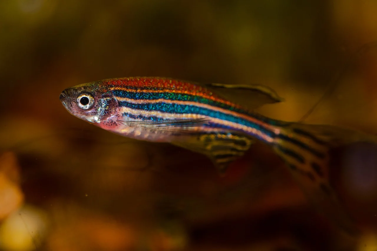
Zebrafish are widely used as vertebrate model organisms, to study the effect of drugs for instance. (Photo: Peter Kuznetsov /Pixabay.com)
“Imagine walking through dense fog. There are objects around you, but you can’t make them out. The objects reflect light though. If you could just filter out the light rays that these objects directly reflect – a tiny fraction of all the light – you would be able to see them. In essence that is the kind of technique we are developing.”
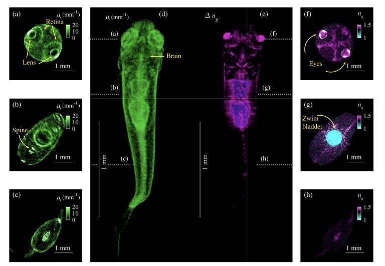
Optical tomography expert, Dr Jeroen Kalkman, and colleagues at the ImPhys/Computational Imaging section (AS Faculty) developed an imaging technique to visualise the inside of zebrafish tissue in high resolution. Kalkman likes to use the fog analogy to describe the technique that shines a light on the inside of this animal.
The findings were recently published in the journal Optica. The article features beautiful high-resolution images of various parts of the zebrafish such as the brain, swim bladder, retina and spine.
‘We want to see how tumours evolve’
The researchers tested their method on dead zebrafish, which they obtained through an ongoing study at Erasmus MC. “But ultimately we should also be able to use it on live fish,” says Kalkman.
And this has a lot of potential. Zebrafish are widely used as vertebrate model organisms, to study the effect of drugs for instance. Often though, the progression of a disease and the effects of medicines entail dissecting the animals at every time step of its progression. With the new technique, Kalkman believes the effects can be monitored while the fish are alive. “We should be able to induce cancer in fish and see how the tumours evolve over time.”
Another application is the analysis of biopsies, small pieces of human tissue that pathologists take from patients for analysis. “Currently, labs often add fluorescent labels to biopsies, or they cut them into small slices and use optical clearing to make them more transparent. This takes a long time and the biopsies can deform during the process. We expect that our technique will be able to image the biopsies in their three-dimensional form without chemical processing, thus helping doctors make more accurate diagnoses.”
Kalkman is now looking for funding to pursue this imaging research. He hopes to collaborate with pathologists and biologists who already have a lot of experience working with tissue biopsies and zebrafish, respectively.

Images from the article ‘Deep-tissue label-free quantitative optical tomography’. (Photos: Jeroen Kalkman)


Comments are closed.