Delft scientists received two grants last week from the Foundation for Fundamental Research on Matter (FOM) for research on the immune system of bacteria and on magnetism.
Animals are not the only creatures suffering from viruses. So do bacteria. These microorganisms have developed a technique to disarm the malignant intruders by chopping virus-DNA into pieces. Better understanding of this process could benefit our battle against our own diseases.
Part of the immune system of bacteria, such as the widespread Escherichia coli, consists of proteins that recognize virus-DNA, bind to it and cut it into bits. Dr. Chirlmin Joo of the bionanoscience group (Faculty of Applied Sciences) and microbiologist Dr. Stan Brouns of the Wageningen University want to find out how these molecules, called CRISPR-associated (Cas) proteins, recognize virus-DNA in the cellular jungle of microbial host DNA. The mechanism behind this process remains elusive.
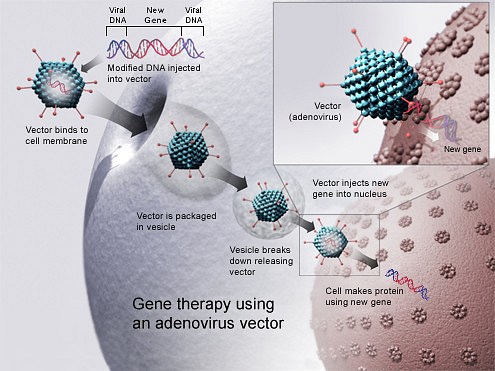
“Insight in the bacterial immune response can be helpful to develop better gene therapy techniques for humans”, said Brouns.
For their project, entitled Deciphering the antiviral genome programming skill of bacteria, the two researchers received over half a million Euros from (FOM).
Joo and Brouns want to study the interaction between the Cas proteins and the virus DNA by labelling the molecules with a fluorescent dye. They will be using a very sensitive light microscope, a ‘total internal reflection fluorescence microscope’.
The grant is part of the subsidy scheme called the Projectruimte, which is created to fund ‘small-scale projects of fundamental research with an innovative character’. 3.3 million Euros have been awarded to a total of eight projects.
Tracing atomic magnets
Dr. Sander Otte of the Kavli Institute of Nanoscience (Faculty of Applied Sciences) also received funding to develop more advanced microscopy techniques.
Otto aims for a better understanding of magnetic materials. “In our lab, we study tiny structures of magnetic atoms by building them from scratch. We build structures atom-by-atom, using low-temperature scanning tunnelling microscopy”, said Otte, who works primarily with cobalt and iron atoms.
“Atoms are placed into their designated positions through atom manipulation, where the apex of the STM tip is used to move them around. Once built, the structures can be probed electronically with atomic precision using the same tip.”
For his proposal ‘Sub-atomic spin resolution through magnetically functionalized scanning probe tips’, Otte received a grant of 400,000 Euros.
Otte compares the tip of his scanning tunnelling microscope with the needle of a turntable. “To that needle we want to attach a little piece of magnetic material. By probing with this tip, we should be able to visualize the field lines surrounding individual atoms.”


Comments are closed.