Herman van Dam (faculty of Applied Sciences) investigated a new method to create a better image of cancer tumours and metastasis. By using a novel PET detector concept, doctors may see three times smaller tumours.
To be able to see better whether a tumour has formed metastases is of crucial importance in finding a way to cure the patient. When cancer is suspected, the patient usually gets a PET scan, which shows where the tumour is located and if there are metastases. At present, this procedure can take a relatively long time – up to 40 minutes – and, moreover, the produced image is often not clear enough, owing to noise and limited spatial resolution. “Because of this, it’s difficult to see if there are metastases, since they’re so small, and it’s not always clear if one sees a metastasis or image noise,” says Herman van Dam.
His new method produces images with higher resolution and larger contrast: “I built several prototypes that can locate the cancer, up to a four times smaller tumour, and do this three times faster than the current method.”
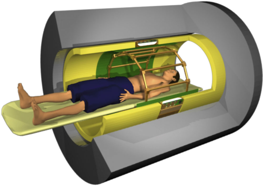
Van Dam focused on the detectors of a PET scanner, the cylinder shaped machine that surrounds the patient and contains radiation detectors, consisting of, in current systems, segmented crystals in the front and a light sensor in the back. Thanks to smart biochemical technology, potential tumours are labelled with so-called tracers, which emit gamma photons. The surrounding detectors use these gamma photons to create a 3D image of the tumours.
Van Dam’s method makes use of one big crystal, instead of segmented ones. And instead of one light sensor, he uses several ones. Since the position of interaction in the big crystal can be determined much more accurately than in the segmented crystals, images with higher resolution can be obtained. One of the crystals Van Dam investigated is called LaBr3:Ce, which was recently discovered at TU Delft.
Van Dam’s supervisor, Dr Dennis Schaart, emphasizes that the new method is a very important step towards creating better images. “It has high promise for the future,” he says. “Fighting cancer is all about having information. By creating a higher resolution image, doctors know better how to deal with the cancer. But it cannot yet be implemented. It has to be calibrated, for example. We already took important steps in that direction. I think we will be able use the method in about five years.”
Van Dam will defend his Ph.D. on 20 March. He is currently a postdoc, working on the follow-up research of the method he developed. In cooperation with Philips, TU Delft is developing a combined MRI and PET scanner. Schaart: “An MRI system works with a magnetic field, and we think we can create a PET scanner that can operate in such a high magnetic field without disturbing the MRI image. Hopefully we’ll be able to create better diagnostic tools.”
De nieuwe tarieven voor het voeren van een rechtszaak (de ‘griffiekosten’) zouden vanaf volgend jaar juli moeten gaan gelden. Het wetsvoorstel is nog niet naar de Tweede Kamer gestuurd, maar minister Opstelten van Justitie heeft het gisteren verspreid onder ‘belanghebbenden’ om hun mening te peilen.
Momenteel zijn de griffiekosten laag voor een bestuurszaak over de studiefinanciering: 41 euro. Studenten zijn meer geld kwijt als ze hun universiteit of hogeschool willen dagen, bijvoorbeeld vanwege een bindend studieadvies of een schorsing: dat kost 152 euro.
De tarieven worden gelijkgetrokken en gaan in principe omhoog: de griffiekosten voor bestuurszaken worden vijfhonderd euro. Maar wie ‘onvermogend’ is, betaalt slechts 125 euro. Waarschijnlijk zijn verreweg de meeste studenten onvermogend. Pas als ze een bijbaan hebben die meer dan 17.300 euro per jaar oplevert, gaan ze extra betalen.
In totaal moet de tariefswijziging de schatkist 240 miljoen euro opleveren. Bijkomend voordeel is dat de tarieven overzichtelijker worden: nu zijn er nogal wat verschillen tussen soorten rechtszaken.


Comments are closed.