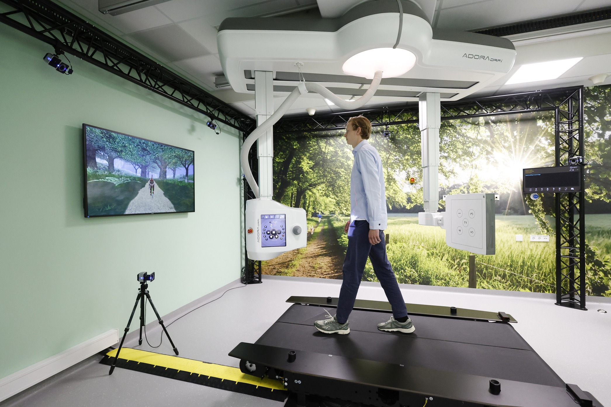
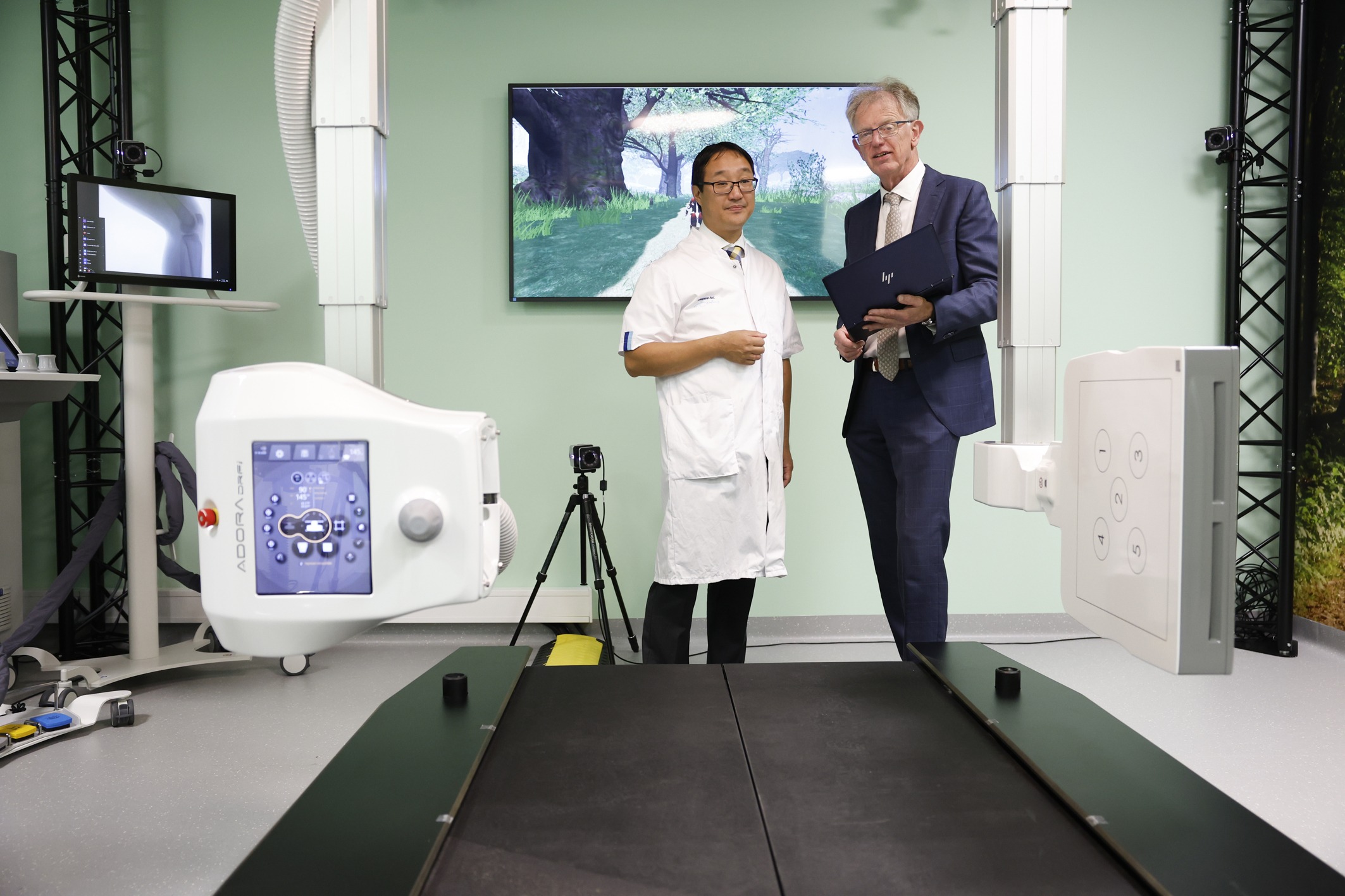
Fifteen seconds of walking on a treadmill surrounded by video cameras and an X-ray camera can reveal the type of osteoarthritis that someone has and the best treatment for it. The Mobi Lab was opened on 17 October by Deans Fred van Keulen (TU Delft) and Stefan Sleijfer (Erasmus MC).

A patient on the treadmill is flimmed with video and X-ray. (Photo: Bas Czerwisnki)
Osteoarthritis is the painful deterioration of joints, such as the knee and hip. It is linked to the loss of cartilage (sometimes referred to as ‘wear and tear’), but an underlying inflammation can also lead to symptoms. Timely and correct treatment may gain some healthy years before a hip or knee replacement is needed. Osteoarthritis affects some one and a half million people in the Netherlands, and this number is expected to rise to 2.3 million by 2040, partly due to the ageing population.
With better diagnosis, it can be determined early on whether osteoarthritis is caused by overuse or inflammation, says Prof. Edwin Oei, Professor of Musculoskeletal Imaging at Erasmus MC. “If the cause is overuse, we move on to supporting insoles, braces or orthopaedic correction (e.g. of bow legs, Eds). But if the symptoms are the result of inflammation, we turn to medication instead.” The Mobi Lab can tell practitioners the degree of excessive joint strain early on.
Lab in hospital
The Mobi Lab has been brought to patients rather than the other way round. The TU Delft MOtion Biomechanics & Imaging (Mobi) Lab, to give it its full name, is located in the middle of Erasmus MC hospital, on the department of Radiology & Nuclear Medicine. The room is painted a soothing green with a wall-sized picture of a forest and birdsong in the background.
The natural ambience is intended to divert attention from the ‘beyond the state of the art’ technology brought together here, as Jaap Harlaar, Professor of Clinical Biomechanics at TU Delft, puts it. A wide treadmill is surrounded by 10 video cameras that capture the gait in spatial detail thanks to small glued-on spheres called reflectors. “But the reflectors slide along the skin which causes a measurement error of a few centimetres,” Harlaar explains. This is why an X-ray camera is also used, which reduces the measurement error to less than a millimetre. The result is a very precise 3D model of the knee joint in motion.
To image cartilage, Oei uses MRI, which may or may not be combined with a PET scan[cheerup-read-more] Positron emission tomography (PET) is an imaging technique that uses radioactive tracers to visualise activity in tissue[/cheerup-read-more]. “On PET scans, the production of bone lights up. That could be a reaction of the bone to the loss of cartilage, which could be an early sign of osteoarthritis,” Oei says about the images. “I get a very detailed image of the cartilage, but I don’t know where the stress is.”
Virtual joint
The 3D computer model of the joint can now be virtually ‘coated’ with a cartilage layer based on the MRI images. The forces affecting the joints are known from treadmill recordings. From these, the pressure distribution within the joint can now be calculated across the entire gait cycle.
“In this way, we hope to find out more about how local overloading leads to osteoarthritis,” says Harlaar. ”Is it the magnitude of the pressure, or does the direction that may create shear stress also play a role? This can now be investigated.”
The Mobi Lab is the first joint facility of the Convergence Health & Technology programme, the research collaboration between TU Delft, Erasmus MC and Erasmus University. Together, the institutions aim to develop solutions to the rising demand and costs of healthcare caused by an ageing population.
Within each step, pressure within the knee joint changes in place and magnitude. (Video: ETH Zürich)
- Also read the Convergence page on the opening programme
Do you have a question or comment about this article?
j.w.wassink@tudelft.nl
Comments are closed.