Knee complaints caused by osteoarthritis are common but poorly understood. Professor Jaap Harlaar develops dynamic imaging for a better understanding and treatment.
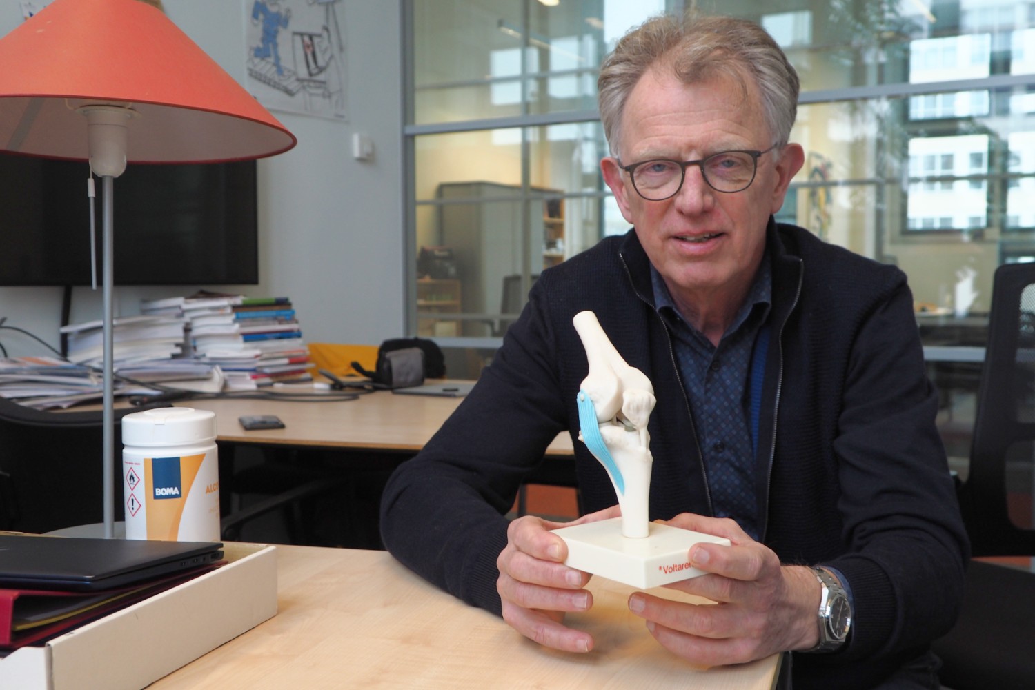
Harlaar wants to provide a better understanding of the moving knee joint. (Photo: Jos Wassink)
- In 2021, nine extra Medical Delta (MD) professors were inaugurated. These professors work at at least two of the five academic institutions represented within Medical Delta (TU Delft, Erasmus MC, Erasmus University, Leiden University and LUMC). The intake brought the total number of MD professors to 22. How does such a double appointment work in practice? That is the underlying question in this miniseries of research portraits.
Arthrosis
According to the thuisarts.nl (in Dutch) website, arthrosis causes the cartilage inside a joint to change. This makes the joint to feel painful and stiff – especially when standing up. Almost one and a half million Dutch people suffer from arthrosis in one or more joints, with knee arthrosis being the most common. Unfortunately, it is not only a disease of old age. Hereditary predisposition, excess weight, overexertion, deviations in standing (e.g. bow legs), or a fall or accident can all lead to accelerated wear and tear of cartilage in the knee, resulting in pain in the joint and the diagnosis of osteoarthritis.
But what causes osteoarthritis symptoms? Where does the wear and tear come from? Where are the thin and worn out patches of cartilage? Why exactly there? And what can be done about it? “With early onset osteoarthritis in young people, it is important to recognise the exact nature of the condition quickly,” says Prof. Jaap Harlaar, Professor of Biomechatronics at the Faculty of Mechanical Engineering, Maritime Technology & Engineering Materials Science (3mE). “Through better characterisation, the treatment team can optimise the treatment and help prevent further damage to the joint.”
Harlaar studied electrical engineering and biomedical engineering at the University of Twente. In April 2017, he was appointed Professor of Clinical Biomechatronics at TU Delft, and became Director of Education for the Clinical Technology (bachelor) and Technical Medicine (master) programmes. In November last year, he also received appointments as Medical Delta Professor at the orthopaedics departments of Erasmus MC and LUMC.
There are various ways to treat osteoarthritis. For example, a knee brace may be prescribed to stabilise the knee or surgery performed to correct a skewed position of the knee. In these cases, the orthopaedic surgeon makes a saw-cut (osteotomy) in the tibia or thighbone and then uses a wedge to move the bone surfaces apart to the desired angle.

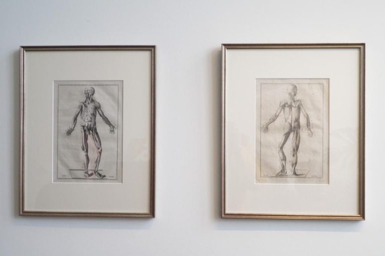
Antique anatomical etches by Eustachius (reprint 1799) in Harlaar’s office. (Photo: Jos Wassink)
Hidden forces
But what goes on inside the knee joint remains well hidden. Harlaar explains that the load on the knee can be as much as two to three times the body weight, and that the force can be as much as four to five times the body weight because of the stabilising muscles and ligaments around the knee. He knows this from the muscle-skeleton computer models that convert measured loads into internal forces on muscles and joints. “But these make a lot of assumptions about the anatomy and about the translation between internal and external movement,” says Harlaar. “For the treatment of individual patients, they still only provide limited information.”
Two techniques are currently available for diagnosing patients: motion capture and ultrasound. The first method, also used in the film industry, captures the movement spatially, but does not provide detailed anatomical information. The second technique provides accurate 3D anatomy, but no motion. As a result, critical questions remain unanswered about where exactly the cartilage is loaded and how (size, direction and duration of forces).
Looking inside the knee
In collaboration with radiologist Edwin Oei, arthrosis researcher Sita Bierma-Zeinstra (Erasmus MC) and biomechanical engineer Bart Kaptein (LUMC), Harlaar is now working on an entirely new form of dynamic screening called biplanar fluoroscopy which should make it clear, through external measurements, what is happening inside the knee. Although the method is still under development, the research project has already been given a catchy name: MOBI (Modelling Osteoarthritis through Biomechanics and Imaging).
Prior to the dynamic measurement, a 3D image of the knee joints is made. During the dynamic measurement, the patient walks on a treadmill at the intersection of two X-ray beams that precisely follow the spatial position of the knee, synchronised with the motion capture. From the combination of pressure, position, and anatomy, biomechanists can calculate the pressure load on the cartilage in the knee.
Harlaar says that “It would be the first time that MOBI would enable orthopaedists to actually look inside the knee to see where the pressure load is occurring and how it is moving through the joint. This should lead to clearer diagnoses and better, more personalised treatment. In the future, it may even be possible to use a digital twin of the patient to assess the effect of a considered treatment.”
Harlaar also expects that MOBI can lead to a better scientific understanding of the origins of osteoarthritis and of the individual differences that are known from practice but so far insufficiently understood.
This is still some way off, but a room has already been reserved for MOBI at Erasmus MC. Harlaar expects the X-ray system to be installed there in the course of 2023. He is also thinking about other applications for MOBI, such as for shoulder joints or backs. “The technique can be applied much more widely. This is an innovation that fits in well with an academic hospital.”
Knee pressure in moving knee joint. Courtesy: Dr. Colin Smith, ETH
Do you have a question or comment about this article?
j.w.wassink@tudelft.nl


Comments are closed.