For the first time scientists recorded neural electricity in mice live. “It’s as if we made a movie of the brain,” says TU researcher and co-author Dr. Daan Brinks.
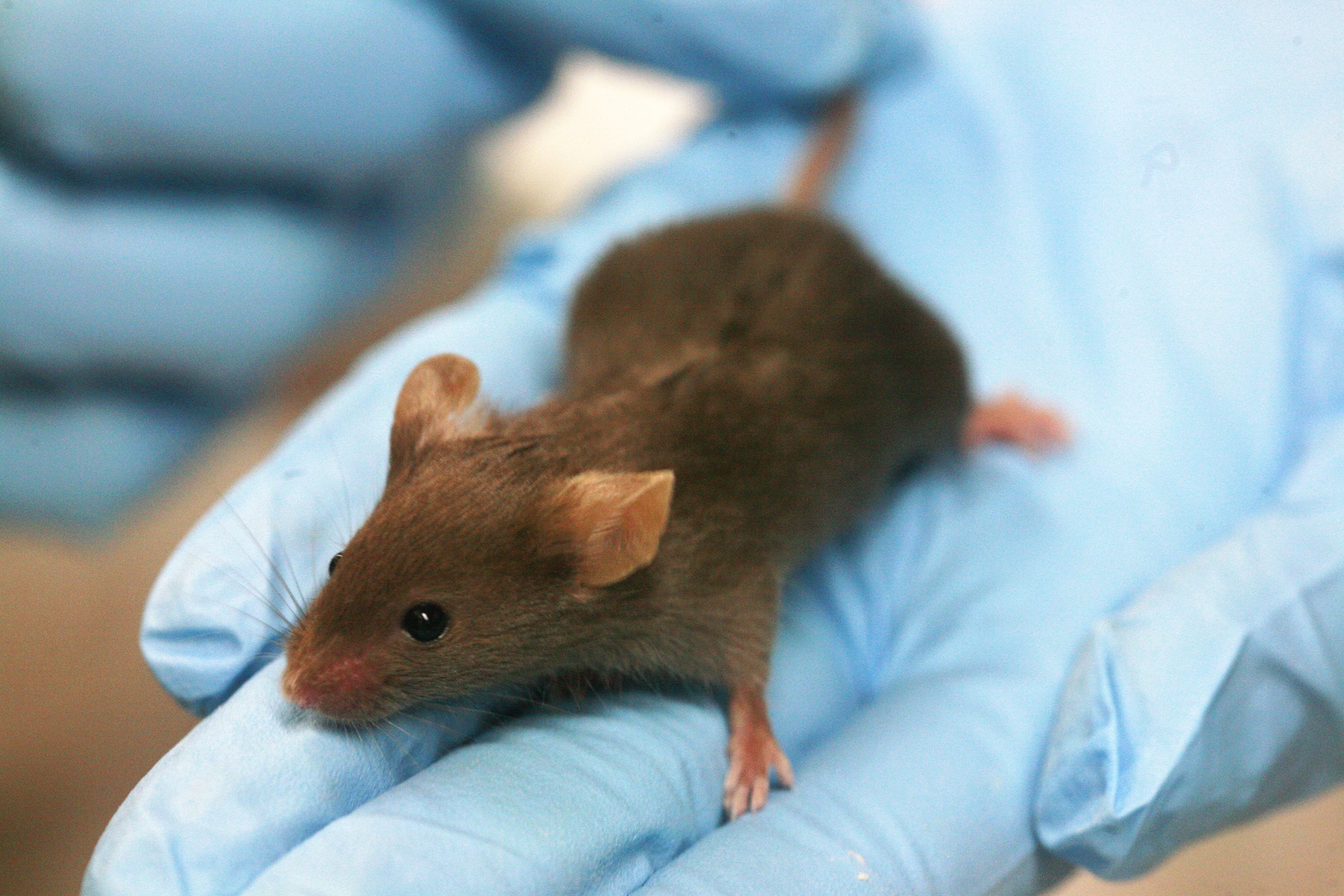
What is he thinking? One day we may find out. (Photo: Wikimedia)
A bunch of genetically engineered mice on a motorised treadmill are at the centre of an important breakthrough in neuroscience. Their skulls were surgically opened, creating windows onto their brains. Researchers, led by the University of Harvard, genetically engineered fluorescent proteins that flash in their brain cells in response to brain activity. They described their work last week in Nature.
Dr Daan Brinks of TU Delft’s Department of Imaging Physics, who until recently worked at Harvard University, is one of the authors of the study. “The brain functions through electrical communication: exquisitely shaped electrical signals carry the information that determine what we think, feel, remember and dream”, he says. “It is known from experiments with electrodes on single cells that a large amount of information about brain function is stored in the precise shape of these electrical signals.”
‘We can really see how electric pulses move’
“It has been possible to measure calcium fluxes in brains for years”, Brinks continues. “But these measurements are an indirect measure of electrical activity. Now we can really see how electric pulses move from cell to cell in a walking, living creature. We could see the animal’s behaviour mirrored in the specific electrical activity in networks of neurons. It is as if we created movies of detailed ‘brain states’ influenced by the animal’s behaviour.”
So how did the researchers get to this feat?
In the 1980s, during an ecological survey of the Dead Sea, an Israeli ecologist found an organism that performs a neat trick: it could convert sunlight into electrical energy in some kind of primitive form of photosynthesis. Decades later, in 2010, researchers at the Massachusetts Institute of Technology (MIT) dusted off the protein, inserted it into a brain, and got the tiny tool to perform its light trick in neurons. When they shone light on the protein-enhanced brain, the tool converted the light into electricity. The researchers could then change the firing of the neurons and, if they chose well, even manipulate the animal’s behaviour.
Adam Cohen, Professor of Chemistry and Chemical Biology and of Physics at Harvard, was intrigued. He wondered if the trick could be reversed. Could the protein convert the electrical activity of neurons into detectable flashes of light? The elusive ‘holy grail’ came after five years of intense, interdisciplinary collaboration between 24 neuroscientists, molecular biologists, biochemists, physicists, computer scientists, and statisticians. First, they tweaked the protein to work in live animals; then they built a new microscope, customised with a video projector.
‘We may find out what memory looks like’
The team is not the first to record neural signals. Hair-thin glass tubes, inserted into brain tissue, can get the job done too. But, these devices record only one or two neurons at a time.
Next, the researchers will make the mouse’s treadmill environment more complex in order to learn more about spatial memory. A sugar station will be added to the set up. Can the mouse remember where to find the sugar station? No one knows what a memory really looks like,” Cohen says in a press release. “Soon, we might.”
In the meantime, the interdisciplinary team will continue to sort through their data, and improve their optical, molecular, and software tools. Better tools could capture more cells, deeper brain regions, and cleaner signals.
Do you have a question or comment about this article?
tomas.vandijk@tudelft.nl


Comments are closed.