A smart combination between optical and electron microscope images single molecules on their way through a living cell.
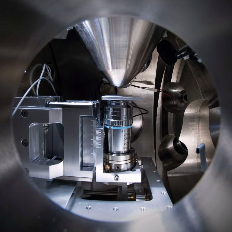
The combination microscope that PhD student Nalan Liv used is basically an add-on to an electron microscope.

In the vacuum chamber, an optical microscope objective has been mounted below and in line with the electron gun of a scanning electron microscope (SEM). Thanks to smart tricks of an electron transparent window (from silicon nitride) and fast calibration procedures, the optical and the electron microscope simultaneously image the same object from top and below. The set-up is known as the Delmic (Delft microscope). Developed by Professor Pieter Kruit and Dr Jacob Hoogenboom, the electron microscope add-on is now the main product of a TU Delft spin-off that started in December 2011.
 Seeing double
Seeing double
Seeing double
A vacuum chamber, caught at the crossroads between an electron beam and a laser, seems an improbable place for life. And yet, imaging life cells and their molecular activities in liquid was exactly what PhD student Nalan Liv set out to do. After her study of biological sciences and bioengineering at the Sabanci University in Turkey, she joined Kruit’s Charged Particle Optics group at TU’s faculty of Applied Sciences in 2009.
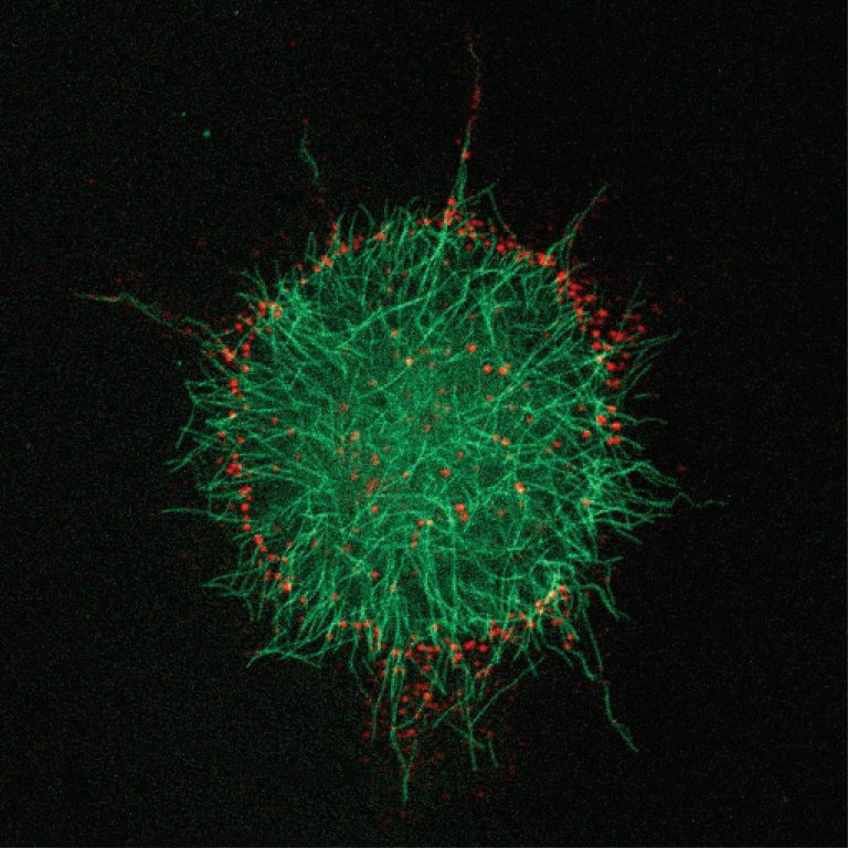
The last chapter in her PhD-thesis, called On-demand electron microscopy of living cells in liquid, shows what the system is capable of: following protein molecules, marked with quantum dots, live on their way through the cell. After being taken up by one of the cell’s antennae (‘filopodial extensions’) the internal transport to the cell’s membrane takes about a minute and a half. Then there is a pause at what Liv calls ‘the docking station’.
Optical fluorescence microscopy will show you the proteins labelled with Q-dots as tiny red points moving around in the dark. Electron microscopy snap shots, taken at any chosen moment, will show you the surrounding cell organelles they interact with.
 Protein tracing
Protein tracing
Protein tracing
At the docking station at the cell’s periphery, the proteins are packed and hitch a ride on the cell’s internal microtubule networks, which transports proteins to the Golgi apparatus where they are processed before being sent to their destination. This internal transport also takes about one to five minutes.
“Sometimes two clusters moving along the tubules collide”, say Liv. “Then there is a short pause during which the cell seems to figure out how to solve the conflict.” A short while later, either the two protein clusters will continue down their own tubule or they will fuse into a larger cluster and go down together.
Correlative microscopy, combining electron and light images, has been developed in several forms since the 1980’s. It usually involves clever tricks to image the same sample subsequently by an optic microscope and an electron microscope. Simultaneous imaging, as the Delmic does, is pretty rare. Certainly when it comes to imaging living cells in liquid, instead of frozen plaques.
Another powerful technique for studying living cells is the superresolution fluorescence microscopy, as developed by TU’s Dr Bernd Rieger. But the required acquisition time makes it hard to follow live action.
Now that proteins, poisons and pharmaceuticals can be traced in living cells, will that revolutionise the way that biologists do science? Well, not overnight, thinks Liv. First of all, electron microscopes are no part of the standard biology lab inventory. Liv thinks that shared facilities may lower the threshold for biologists to access combination microscopy.
Besides, researchers tend to cling to the method they have learned to master. Optical fluorescence microscopy and electron microscopy have their own separate ways of preparing and labelling samples. People don’t like to change that, Liv observes. To convince them, it’s not enough to read about some new microscopy technique. “People will want to see it work for their own experiments.” And that is where the shared facilities may play a role.
“Pieter Kruit’s technique will be appreciated later”, Liv expects. “In a couple of years.”
–> Nalan Liv, Simultaneous Correlative Light & Electron Microscopy of Samples in Liquid, PhD-thesis supervisors Prof. Pieter Kruit and Dr Jacob Hoogenboom, defence 03 October 2014.
Do you have a question or comment about this article?
delta@tudelft.nl
Comments are closed.