When scanning a lab mouse, its posture in the scanner is a bit different every next time. That makes it hard to follow the biological processes. PhD student Peter Kok developed a method that maps different images into a common frame.
A common frame of reference helps to trace the development a particular animal through time. The procedure that Peter Kok developed combines the computer tomography (CT) data with a standard 3D atlas of the mouse skeleton.

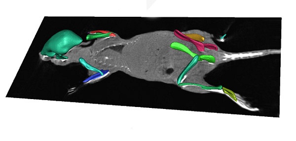
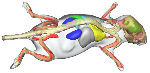
After a course alignment of the entire skeleton, individual bones are registered step-by-step in a process that starts with the skull, proceeds with the spine and then moves down the front and hind limbs separately. Then it scans the lungs, skin, and other organs. The result looks like this.
Next, each bone gets a bounding box (yellow in the image below). A computer algorithm resamples the original CT data to obtain a volume mapped to the standardized bone.
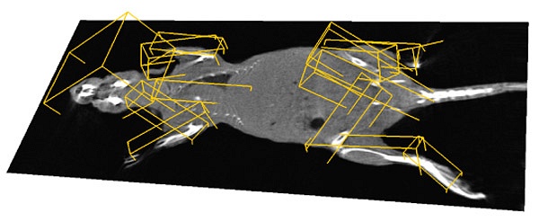
In order to prevent different elements from overlapping, the user can move separate elements apart to get a better view. The user may select a camera point of view for the best angle.
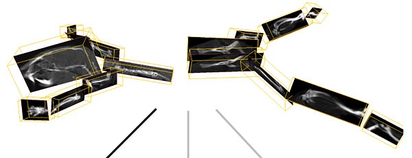
Once the data are mapped to a standard reference, researchers can follow physiological processes in time by comparing CT scans form different points in time of the same animal. Here, for example, the left side of the femur bone gets resorbed at the place where a tumor grows.

Ideally, the image processing should accommodate different modes of imaging like computer tomography (CT), magnetic resonance imaging (MRI) and bioluminescent imaging (BLI). Dr. Peter Kok says over the telephone that MRI and BLI data may be mapped onto the standard mouse atlas, just like CT data. Indeed, different imaging modes can be combined to add anatomical information (CT) to tumor localization (by BLI).
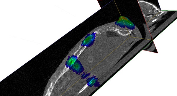
For areas of special interest, said Kok, even microscopy images may be fitted onto the same frame of reference by registering the image with two or more recognizable landmarks.
Thus, Kok has developed a powerful tool that allows integration and comparison of lab mouse images. A researcher can follow one specific animal through time over scales varying from microscopic to whole body scans. Or images of different animals may be matched for comparison.
Nonetheless, Kok expects that his software will eventually be redundant, according to proposition number 7 in his thesis: ‘All animal testing will be replaced by simulations in systems biology’.
Let’s hope his proposition works out quickly.
–> Peter Kok, Integrative visualization of whole body molecular imaging data, December 12 2014, PhD supervisor Prof. Boudewijn Lelieveldt.


Comments are closed.