When building a cell from scratch, one of the challenges is to make it divide. Nanobiologist Elisa Godino recreated mechanisms inspired from bacteria in artificial cells.
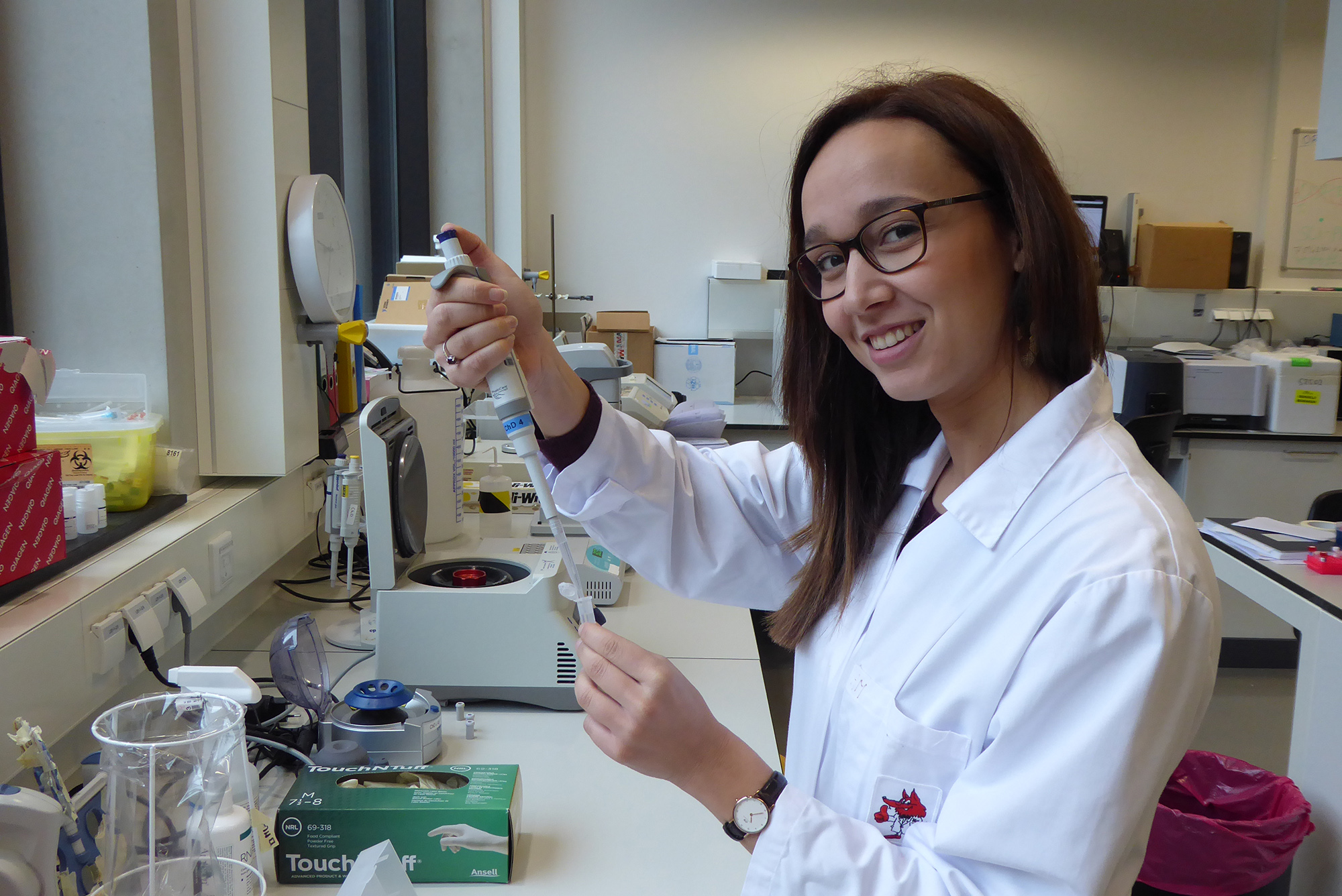
Elisa Godino has a fascination for artificial life. (Photo: Jos Wassink)
Cell division is a basic vital function. Without it, hairs wouldn’t grow, your bowels couldn’t work, and organisms couldn’t reproduce. Without cell division, life would basically stop in its tracks. Cell division is one of life’s basic processes that needs to be mastered in the quest for artificial life.
When studying biotechnology in Trento, Italy, Elisa Godino worked on protocells – autonomous cell-like structures exhibiting genetically controlled behaviour and metabolic energy production. She developed a fascination for synthetic life, creating life from inanimate materials, and she contacted Professor Christophe Danelon for a PhD position, which started January 2018.
Creating artificial life, or making a living synthetic cell, is an exciting international field of research in which rapid progress is being made. The first Dutch symposium on Building a Synthetic Cell (BaSyC) took place in Delft in 2018.
Different groups work on various topics such as making the cell membrane out of a lipid bilayer, storage and retrieval of information in the cell’s DNA including the production of functional proteins. Plus the self-organisation of vital functions through active proteins.
The research by Elisa Godino and colleagues at the Kavli Institute of Nanoscience (TU Delft Faculty of Applied Sciences, Bionanoscience department) falls in the domain of protein-mediated self-organisation of the cell, focussing on cell division.
Finding the middle
When a cell divides, it splits neatly in half. Nanobiologists have long wondered how cells achieve that. How does a cell even know where its middle is? Professor Cees Dekker started the research into cellular geometry in 2015. He and his postdoc experimented with proteins known to play a pivotal role in mid-cell positioning. Meet the Min-proteins.
Min proteins oscillate between the poles of a bacterium, no matter what shape it takes, Dekker’s team showed. The net effect of the Min-proteins migrating between the poles is that their average concentration is lowest in the middle of the cell. And that’s how a bacterium ‘knows’ where its middle is.
Four years on, researchers in Professor Christophe Danelon’s lab are replicating the bacterial cell division mechanism in artificial ‘cells’. These proto-cells consist of vesicles less than 10 micrometres across, surrounded by a lipid double-layer. The micro-bubbles contain DNA that codes for Min-proteins and also the cellular machinery called PURE. This molecular machine reads DNA, transcribes it into RNA, and synthesises the associated protein. This process starts automatically as soon as the ‘cell’ is warmed up. Elisa Godino and colleagues reported on their results in Nature Communications.
Pulsating bubbles
Under the microscope, researchers saw how the internally produced Min-proteins and a green-marked reporter protein migrated periodically between the vesicle’s interior and the membrane. They also noticed a change in the bubbles’ shape. Bubbles spontaneously changed from circular (Min-proteins in solution) to elongated (Min-proteins on membrane) and back.
“We observe an autonomous deformation of liposomes driven by internal Min-oscillations”, says Danelon.
As said, a bacterium uses Min-protein oscillations to determine its middle (where the concentration is lowest). But another protein, called FtsZ, forms a chain that constricts the cell membrane to the point where half of it buds off.
“We have now shown the localisation mechanism”, says Godino. “The next step is to combine the localisation with the formation of a ring that divides the cell.” And, with a sudden smile, “I still have another two years to go.”
- Elisa Godino, Jonás Noguera López, David Foschepoth, Céline Cleij, Anne Doerr, Clara Ferrer Castellà & Christophe Danelon, De novo synthesized Min proteins drive oscillatory liposome deformation and regulate FtsA-FtsZ cytoskeletal patterns, Nature Communications, 31 October 2019.
Do you have a question or comment about this article?
j.w.wassink@tudelft.nl


Comments are closed.