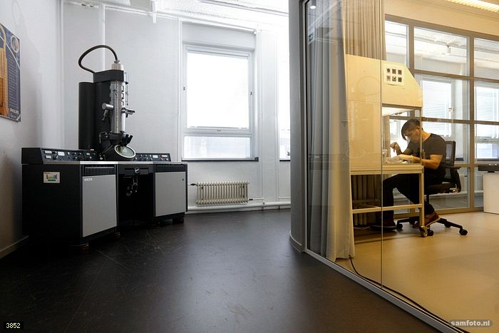
In search of TU Delft’s heritage, Delta discoverd a 50-year old electron microscope in the corner of the new glass VLLAIR microscopy laboratory: the Philips EM300.
What first draws the eye is the fluorescent green screen directly beneath the electron column. Electrons that pass through the preparation form an enlarged image on the screen. Researchers would examine the image via the optical microscope, which is attached to the outer side due to the vacuum in the microscope.
Details smaller than 1/10th of a nanometer
According to microscopy expert Prof. Pieter de Kruit, from the faculty of Applied Sciences, it is difficult to overestimate the significance of electron microscopy. The ability to form an image of details smaller than 1/10th of a nanometer using the De Broglie wavelength of accelerated electrons has made the atomic world visible, so we can now describe the internal structure of cells and bacteria. The electron microscope has also become an essential tool in materials science for studying the connection between structures and macroscopic properties such as strength and rigidity. Furthermore, electronic circuits could never have been miniaturised without the electron microscope.
Designed and built by a student of TU Delft
The first electron microscope was constructed in 1931 by Ernst Ruska, who went on to receive the Nobel Prize, at the Technical University of Berlin. Delft Physics Professor Hendrik Dorgelo was close to Ruska but had no budget to purchase one of his microscopes. In 1939, Dorgelo’s student Jan Bart Le Poole proposed to design and build an electron microscope for his graduation project. Despite the intervention of the Second World War, Le Poole succeeded in creating his first image by 8 April 1941. A year later, he began constructing a 150 kilovolt transmission electron microscope at the Technical Physics Department at the TNO/Delft Institute of Technology, financed by the Delft University Fund and several Dutch companies. The war forced Le Poole to be self-reliant. Nevertheless he contacted Philips, believing that the electron microscope could be of interest to the company.
Many notable figures came to see it
Following the war, Le Poole caused a sensation with demonstrations of his microscope. Many notable figures came to see it, including Queen Juliana and Prince Bernhard. Thanks to various important personages personally urging Anton Philips to reconsider, Philips eventually manufactured a series of electron microscopes. It established Philips Electron Optics, with Le Poole, who had since become Professor of Electron Optics at the Delft Institute of Technology, in an advisory role. The first model of electron microscope was the Philips EM100; the company sold 400 of these. In 1967 the EM300 followed, with a more sophisticated focusing device and the ability to produce diffraction patterns for identifying the structure of micro-crystals. Now, 50 years on, the old workhorse that revealed viruses, crystals, organelles and so many other wonders of the atomic world to researchers stands in a corner as a piece of history.
Source: P. Kruit, ‘De Philips EM300 Elektronenmicroscoop’, 175 jaar TU Delft, Erfgoed in 33 verhalen, published by Histechnica, 2017


Comments are closed.